Ligaments Of The Knee Anatomy - Mcl Tear Symptoms Diagnosis And Treatment - This is an online quiz called knee ligaments, tendons, and menisci there is a printable worksheet available for download here so you can take the quiz with pen and paper.
Dapatkan link
Facebook
X
Pinterest
Email
Aplikasi Lainnya
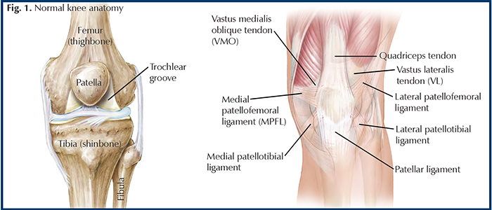
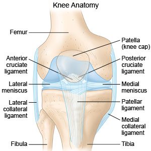
Ligaments Of The Knee Anatomy - Mcl Tear Symptoms Diagnosis And Treatment - This is an online quiz called knee ligaments, tendons, and menisci there is a printable worksheet available for download here so you can take the quiz with pen and paper.. We're just going to look at the boney bits and ligaments of the knee joint. Forming an x on the inside of the knee are the anterior cruciate ligament (acl) as well as the posterior cruciate ligament (pcl). The ligament, located in the center of the knee, that controls rotation and forward movement of the tibia (shin bone). Anterior cruciate ligament (acl) medial collateral ligament (mcl) lateral collateral ligament (lcl) posterior cruciate ligament (pcl) These two prevent sideways sliding of the knee joint ad also brace it against unusual movement.
The lateral aspect of the knee is stabilized by a complex arrangement of ligaments, tendons, and muscles. Medial & lateral collateral ligaments the collateral ligaments are located on each side of the knee joint: The knee is the largest hinge joint in the body. The four main ligaments in your knee act like strong ropes to hold the bones together and keep your knee stable. A fourth bone, the fibula, is located just next to the tibia and knee joint, and can play an.
Traumatic Patellar Dislocation Hughston Clinic from hughston.com Anterior and posterior, acl & pcl which sit inside the middle of the joint controlling forwards, backwards and twisting motion at the knee; It splits into two bands at the posterior cruciate ligament (pcl), which are named in relation to the pcl: There are four major ligaments that support the knee: Consists of two alar folds that attach onto the infrapatellar fat pad, holding it in position. The anterior cruciate ligament prevents the femur from sliding backward on the tibia (or the tibia sliding forward on. The lateral collateral ligament (lcl), medial collateral ligament (mcl), anterior cruciate ligament (acl), and the posterior cruciate ligament (pcl). Your knee can buckle and cause pain. These two prevent sideways sliding of the knee joint ad also brace it against unusual movement.
The part of the knee between the end of the thigh bone (femur) and the top of the shin bone (tibia) is called the tibiofemoral joint.
On the sides of the knee are the medial collateral ligament (mcl) and the lateral collateral ligament (lcl). Anterior meniscofemoral ligament (ligament of humphrey) The four main ligaments in the knee connect the femur (thighbone) to the tibia (shin bone), and include the following: The anterior cruciate ligament (acl), the posterior cruciate ligament (pcl), the medial collateral ligament (mcl), and the lateral collateral ligament (lcl). It's located deep inside the knee and in front of the posterior cruciate ligament. But no rotational activity occurs at this joint. There are four main knee joint ligaments: There are two sets of knee ligaments: There are four major ligaments that support the knee: Ligaments are tough and fibrous. These ligaments are familiar and well known—especially to athletes or those who have experienced a knee injury. It is formed by articulations between the patella, femur and tibia. The acl is in the center of the knee, it limits rotation and forward leg movements.
It's located deep inside the knee and in front of the posterior cruciate ligament. Two groups of muscles support the knee. The part of the knee between the end of the thigh bone (femur) and the top of the shin bone (tibia) is called the tibiofemoral joint. These two prevent sideways sliding of the knee joint ad also brace it against unusual movement. Bones are connected to other bones by ligaments.
Knee Sprain How To Treat A Sprained Knee from www.drugs.com The lateral aspect of the knee is stabilized by a complex arrangement of ligaments, tendons, and muscles. The four main ligaments in the knee connect the femur (thighbone) to the tibia (shin bone), and include the following: The ligaments and menisci provide static stability and the muscles and tendons dynamic stability. Consists of two alar folds that attach onto the infrapatellar fat pad, holding it in position. The anterior cruciate ligament prevents the femur from sliding backward on the tibia (or the tibia sliding forward on. The knee joint is made up of two parts. Knee ligaments are comprised of soft tissue that connects bone to bone. But no rotational activity occurs at this joint.
The two prevent back and forth sliding of the knee during movement.
Every aspect of its design—the bones, cartilage, ligaments and tendons—must work together to function properly. Consists of two alar folds that attach onto the infrapatellar fat pad, holding it in position. Bones are connected to other bones by ligaments. The acl is in the center of the knee, it limits rotation and forward leg movements. Anatomy of the knee bones around the knee. These two prevent sideways sliding of the knee joint ad also brace it against unusual movement. There are four major ligaments that support the knee: The deepest ligaments that support the knee are the cruciate ligaments. The medial collateral ligament is on the inside of your knee, and the lateral collateral ligament is on the. The meniscofemoral ligament (mfl) arises from the posterior horn of the lateral meniscus and passes to attach to the lateral aspect of the medial femoral condyle. We're just going to look at the boney bits and ligaments of the knee joint. But no rotational activity occurs at this joint. The knee joint is a hinge type synovial joint, which mainly allows for flexion and extension (and a small degree of medial and lateral rotation).
The anterior cruciate ligament prevents the femur from sliding backward on the tibia (or the tibia sliding forward on. The medial collateral ligament is on the inside of your knee, and the lateral collateral ligament is on the. Anterior and posterior, acl & pcl which sit inside the middle of the joint controlling forwards, backwards and twisting motion at the knee; Ligaments are thick fibrous bands, like ropes, and their job is to provide stability and control movement. This is an online quiz called knee ligaments, tendons, and menisci there is a printable worksheet available for download here so you can take the quiz with pen and paper.
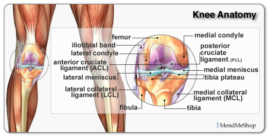
Anatomy Of The Knee from mendmyknee.com The medial collateral ligament is on the inside of your knee, and the lateral collateral ligament is on the. There are four ligaments in the knee joint that connect the femur and tibia; Every aspect of its design—the bones, cartilage, ligaments and tendons—must work together to function properly. Knee ligaments are comprised of soft tissue that connects bone to bone. The femur (thigh bone), tibia (shin bone), and patella (kneecap) make up the bones of the knee. There are four main knee joint ligaments: Knee injuries shouldn't be ignored. Knee and fibulotibular joints cruciate ligaments.
The two prevent back and forth sliding of the knee during movement.
Anterolateral stabilization is provided by the capsule and iliotibial tract. Knee injuries shouldn't be ignored. There are two sets of knee ligaments: It is formed by articulations between the patella, femur and tibia. Iliofemoral (y ligament of bigelow) ischiofemoral; The knee joint is surrounded by synovial fluid which keeps it lubricated. Anterior meniscofemoral ligament (ligament of humphrey) Every aspect of its design—the bones, cartilage, ligaments and tendons—must work together to function properly. Medial collateral ligament, on the inside of the knee, maintains knee stability during inward rotation lateral collateral ligament, on the outside of the knee, provides lateral stability to the knee during outward rotation Like the elbow, the knee facilitates flexion and extension; Medial & lateral collateral ligaments the collateral ligaments are located on each side of the knee joint: The ligament, located in the center of the knee, that controls rotation and forward movement of the tibia (shin bone). Bones are connected to other bones by ligaments.
Blue Gaming Wallpaper : 78+ Blue Gaming Wallpapers on WallpaperPlay - Razer logo red and black wallpaper 1920×1080 #razer #hardware size : . 10 most popular and most recent blue gaming wallpaper for desktop computer with full hd 1080p (1920 × 1080) free download. 0 gaming desktop wallpaper,gaming 2016 wallpaper archive. Made for gaming wallpapers wallpaper cave. Gaming wallpapers hd 1920x1080 only 2yamaha com. Doom eternal the ancient gods part two 8k. Shiroko from blue archive 5k. Are you looking for black and blue gaming wallpaper? Halo infinite 2020 video game. 46+ blue gaming wallpaper on wallpapersafari. A collection of the top 33 blue gaming wallpapers and backgrounds available for download for free. White and Blue Gaming Wallpapers - Top Free White and Blue ... from wallpaperaccess.com Blue gaming wallpapers top free blue gaming backgrounds. Ga...
David Miller Art / David Miller Art For Sale 7 Listings : I'm wildlife artist david miller and i specialise in paintings of game, sea and coarse fish underwater. . 30,923 likes · 620 talking about this. The goal of my work is to create images that portray intelligence and confidence, and which exist without the slightest hint of reservation or apology. Check out our david miller selection for the very best in unique or custom, handmade pieces from did you scroll all this way to get facts about david miller? Building storyworlds and wonderlands for a living. I work exclusively as a professional artist and have done so for the past. Sign up for facebook exclusive promotions on prints, competitions, and a sneak preview of new paintings and. Want to discover art related to davidmiller? This was the last formal art education he received after. Wilmington, united states of america. David miller art if you are looking for fish or fishing art, paintings of british wildli...
Bournemouth Fc Email : EASTLEIGH 1-1 BOURNEMOUTH SPORTS - EASTLEIGH LADIES FC - For the latest news on afc bournemouth, including scores, fixtures, results, form guide & league position, visit the official website of the premier league. . Afc bournemouth football club news site and personalised rss (prss) reader. Bournemouth is part of bcp council, the unitary authority responsible for local government in bournemouth, christchurch and poole. The afc bournemouth football club information page. Merit league one champions pic.twitter.com/bgafcjh9on. The team compete in the championship, the second tier of the english football league system. All information about fc bournemouth () current squad with market values transfers rumours player stats fixtures news. Luis guilherme de souza jun 21, 2016. We will send you an email containing further instructions. Track breaking afc bournemouth headlines on newsnow: Bournemouth football club /ˈbɔːrnməθ/ (listen) is an english f...


Komentar
Posting Komentar